Vertigo Neurology
SYMPTOM OVERVIEW
• Vertigo: a condition that is usually due to a disturbance or asymmetry in the vestibular system involving the semicircular canals or the central nervous system structures.
• Key anatomical components: semicircular canals (horizontal, posterior; superior); otolithic apparatus (utricle and saccule)
• Types of vertigo: Peripheral (80% of cases) and Central
• Peripheral vertigo: benign paroxysmal positional vertigo; Meniere's disease; Cogan's syndrome (see MMW link); otitis media; vestibular neuritis; acoustic neuroma; semicircular canal dehiscence syndrome; perilymphatic fistula; aminoglycoside toxicity; recurrent vestibulopathy; herpes zoster oticus; and labyrinthine concussion
• Central vertigo: multiple sclerosis; cerebellar infarction and hemorrhage; brainstem ischemia; migrainous vertigo; chiari malformation;
episodic ataxia type 2

SIGNS AND SYMPTOMS
• Nystagmus: a rhythmical oscillation of the eyeballs
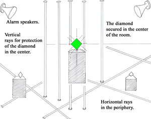
o Horizontal: usually occurs in peripheral vertigo as illustrated by the horizontal security light beams surrounding the periphery of the diamond. However, horizontal nystagmus is occasionally noted in patients with poor pursuit due to large central lesions.
♣ Peripheral lesions: fast component away from affected side; visual fixation suppresses nystagmus (robber becomes focused on the diamond while standing in the periphery, thereby, stopping the nystagmus)
o Vertical (upbeat/downbeat): usually occurs in central vertigo as illustrated by the vertical security light beams shinning down on the central aspect of the diamond. However, central vertigo may present with various patterns of nystagmus.
• Central lesions: Nystagmus may reverse direction with convergence (convergence of vertical light beam on diamond)

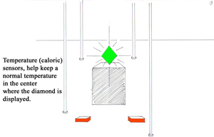
• Caloric testing: Lack of a response to warm or cold water on one side suggests disease on that side, usually in peripheral lesions.

• Postural Instability
o Peripheral lesions: walking is usually preserved
o Central lesions: frequent falls occur while walking
• Auditory
o Peripheral lesions: deafness, tinnitus, or ear pain may occur
o Central lesions: rarely have auditory complaints

DIAGNOSIS
• Dix-Hallpike maneuver: useful for the confirmation of benign paroxysmal positional vertigo
• Audiometry: useful in the evaluation of peripheral lesions and in the confirmation of Meniere's disease.
• Imaging: MRI/MRA are useful in evaluation of central lesions. MRI/CT are useful in evaluating acute sustained vertigo in some circumstances.
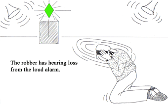
The Tulio Phenomenon illustrated here consists of sound induced vertigo or nystagmus that can be caused by superior canal dehiscence; perilymphatic fistula; and Meniere's disease.
• A peripheral lesion in the inner ear may present with deafness and tinnitus.

TREATMENT
• Treatment of the underlying cause
• Symptomatic treatment duration of 48hrs or less with one of the following drug classes: anticholinergics; antihistamines; benzodiazepines; antiemetics observing potential drug –drug interactions
• Vestibular exercises/rehabilitation
- Anatomy
- Anesthesiology
- Cardiovascular Medicine
- Dermatology
- Emergency Medicine
- Endocrinology
- Gastroenterology
- Geriatric Medicine
- Gynecology
- HIV
- Hematology and Oncology
- Infectious Disease
- Internal Medicine
- Nephrology
- Neurology
- Obstetrics
- Ophthalmology
- Orthopedics
- Otolaryngology
- Pediatrics
- Pharmacology
- Prevention
- Psychiatry
- Pulmonology
- Radiology
- Rheumatology
- Surgery
- Urology
- case report
- trial reference


